

When a joint problem persists, a provider often needs a clear view of the structures beneath the surface to better understand the cause. Magnetic resonance imaging (MRI) offers a detailed way to examine a joint, and it doesn’t require surgery. This tool helps reveal soft tissue structures that are not visible on standard imaging. Clinicians can use the information to support diagnosis, guide treatment decisions, and better understand what may be contributing to ongoing symptoms. Here’s more information about MRI and how they help diagnose joint disorders:
Understanding the Imaging Process
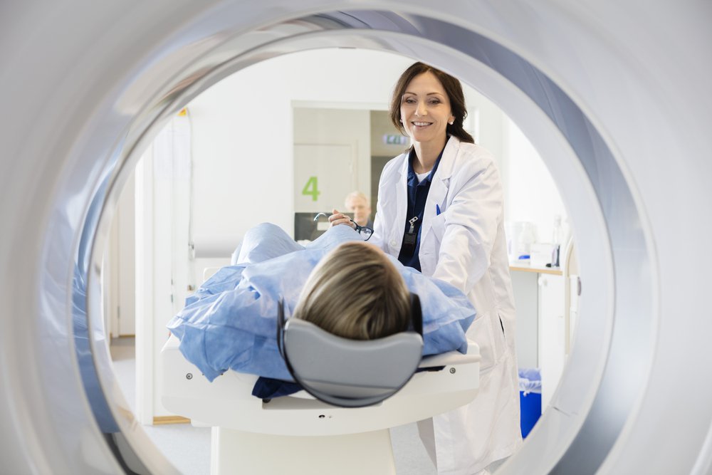
An MRI produces detailed images of the inside of the body by using a strong magnetic field and radio waves rather than ionizing radiation. During the scan, you lie still on a table that slides into the imaging unit. The machine collects signals from the tissues being examined. A computer then processes those signals and converts them into cross-sectional images. These images are taken from multiple angles; this allows providers to evaluate the joint in greater detail. Because the procedure is noninvasive, it offers a clear view of internal structures without the need for surgical incisions or extensive recovery steps.
Examining the Joint Structures
The strength of an MRI lies in how clearly it shows soft tissue structures. Many joint conditions affect areas that other imaging methods may not capture well, and MRI provides significantly more detail. This makes it especially useful when a provider needs information beyond standard bone imaging, because it reveals structures that are otherwise difficult to evaluate.
An MRI of a joint may show several types of structures, including:
- Cartilage: The smooth tissue that cushions the ends of bones within the joint
- Ligaments: The bands that connect bone to bone and support stability
- Tendons: The tissue that attaches muscle to bone around the joint
- Bone: The underlying structure, including internal changes within the bone
- Soft tissue and fluid: Surrounding material and any buildup in the area
By showing these structures in detail, MRI enables a provider to locate the source of a problem. It also helps assess how extensive the condition may be, which supports clearer decision-making. This clarity offers a more complete view than a single image alone, and it helps guide appropriate next steps in care.
Applying the Diagnostic Findings
A provider may interpret the MRI images alongside your medical history, physical examination, and other relevant diagnostic tests. These combined elements provide a more comprehensive understanding of the joint and the factors contributing to symptoms, and they support more informed clinical decision-making. The imaging results then guide the next steps in the diagnostic process, as the provider reviews structural details, compares them with reported symptoms, and assesses how well the findings align with the overall presentation. This structured evaluation helps narrow diagnostic possibilities and supports the development of an appropriate treatment plan.
MRI findings also function as a key reference point over time. If a provider needs to monitor changes within a joint, the original images establish a reliable baseline for comparison. With that record in place, the provider can track progression more accurately; they’re able to adjust the care approach as new information becomes available.
Learn More About MRI
An MRI provides a detailed, noninvasive method for examining the internal structures of a joint, including cartilage, ligaments, and tendons. Providers interpret these images alongside your medical history and physical examination. This helps create a more complete understanding of a joint disorder, and it supports the diagnostic process. If you have a joint concern requiring further evaluation, ask your provider whether an MRI is appropriate for your situation.
