

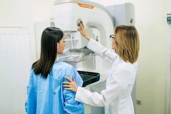
When a doctor finds a lump or an unusual spot on a mammogram, the next step often involves breast pathology. Breast pathology is the study of breast tissue and cells under a microscope. A pathologist examines samples from a biopsy or surgery, and the findings shape your next steps with your care team. Here is more information about this process to help you prepare for your results:
Diagnosing Breast Cancer
A biopsy gives the pathologist a small piece of tissue to study. Before anyone looks at it under a microscope, the lab preserves the tissue and slices it very thin. Technicians stain the slices with dyes, and the dyes highlight the structure of each cell.
The pathologist compares your cells to normal breast tissue, and differences in size or shape stand out. Cancer cells look crowded and irregular. If the cells appear normal, the breast pathology report may describe the area as benign.
Some results fall into a gray zone because the cells look atypical but not cancerous. In these cases, the pathologist may order extra stains, or a second expert may review the slides. You may ask your doctor for a copy of the full report.
Classifying Tumors
Not all breast cancers behave the same way, and the pathologist identifies the specific type. Classification depends on where the cancer started and whether it has spread into nearby tissue. Your report will likely name one of the following types.
- Invasive ductal carcinoma: Cancer spreads beyond the ducts into nearby tissue.
- Invasive lobular carcinoma: Cancer starts in the milk-producing glands and spreads.
- Inflammatory breast cancer: A rare type that blocks lymph vessels in the skin.
After the pathologist identifies the type, the lab tests the tumor for certain proteins. These include estrogen receptors, progesterone receptors, and HER2. Hormone receptors respond to estrogen or progesterone, and HER2 affects cell growth.
Grading Aggressiveness
Grading describes the appearance and growth pattern of cancer cells. Pathologists assign a grade from 1 to 3, and each number reflects a different level of change. A grade 1 tumor has cells that look closer to normal tissue. Grade 3 cells look very different and divide quickly.
Since grade relates to growth speed, your care team uses it alongside stage and receptor status. Grade and stage are different measures. Stage describes tumor size and spread, but grade focuses on the cells themselves. You may want to ask about both numbers at your next appointment.
Guiding Treatment Courses
Your oncologist reads the pathology report closely, and its details help shape a treatment plan. Each finding points toward certain treatment options. When your team plans treatment, they typically review these details:
- Tumor type and size
- Grade
- Hormone receptor status
- HER2 status
- Margins, or the distance between the tumor and the edge of removed tissue
- Lymph node involvement
Margin results matter after surgery. If cancer cells reach the edge of the removed tissue, your surgeon may recommend another procedure. Patients with HER2-positive cancers often receive targeted drugs, and hormone-positive cancers may call for hormone therapy. Your care team can explain how each result affects your options.
Schedule Breast Pathology Services
If your doctor has ordered a biopsy, our team can process and review your tissue samples. A pathologist can prepare detailed reports, and they send results directly to your physician. Contact an office with questions about timing or next steps. Call a clinic today to schedule services or learn more about the process.
