

Breast cancer affects millions of people each year, and early detection helps people get the care they need. A breast ultrasound is one of the tools doctors use to examine breast tissue, identify abnormalities, and guide next steps in care. Alongside other imaging methods, it gives physicians a clearer picture of your breast health. Here is more information about breast ultrasound and its benefits:
Understanding Breast Ultrasound
A breast ultrasound uses high-frequency sound waves to create images of the tissue inside the breast. The sound waves bounce off tissue and return to a transducer, which converts them into real-time images on a screen. Doctors use ultrasound to examine lumps found during a physical exam, and it can distinguish between solid masses and fluid-filled cysts. Solid masses require further evaluation because they may be cancerous, while cysts are generally benign.
Exploring the Procedure
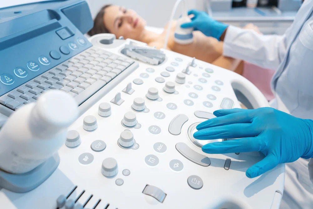
The procedure is straightforward, and it may be more comfortable for the patient than a mammogram. A technologist applies a clear, water-based gel to the breast, which helps the transducer glide smoothly across the skin. The technologist then moves the transducer across the area of concern, capturing images from multiple angles.
The appointment typically lasts between 10 and 15 minutes, but this varies depending on the complexity of the scan. No radiation is involved. While mammograms can be uncomfortable, this procedure is painless for most patients.
Reading the Results
After the scan, a radiologist reviews the images and prepares a report for the referring physician. Radiologists classify findings using a standardized system called BI-RADS, or the Breast Imaging Reporting and Data System. This system uses ratings from zero to six:
- Category zero: Incomplete; more imaging is needed
- Category one and two: Negative or benign findings; routine follow-up is recommended
- Category three: Probably benign; short-term follow-up is suggested
- Category four and five: Suspicious or highly suggestive of malignancy; biopsy may be recommended
- Category six: Known malignancy already confirmed by biopsy
The referring physician reviews the radiologist’s report and discusses the findings with the patient. A higher BI-RADS category does not confirm cancer, but it may indicate that further testing is warranted.
Comparing Breast Ultrasound and Mammogram
Mammograms remain the standard screening tool for breast cancer, and ultrasound is not a replacement. The two tools serve different purposes and work together to detect abnormalities. Mammograms use X-ray technology and are well-suited for detecting calcifications, which can be early signs of cancer. Ultrasound is particularly useful for people with dense breast tissue because ultrasound is more sensitive in detection than X-rays in dense tissue.
Ultrasound also has advantages in specific situations:
- It does not use radiation, making it a safer option for pregnant patients
- It provides real-time imaging, which is useful for guiding biopsies
- It can clearly differentiate between solid masses and fluid-filled cysts, which mammography cannot always do
For these reasons, physicians may recommend ultrasound as a supplemental tool when a mammogram produces unclear results or when dense tissue limits visibility.
Learn More About Breast Ultrasound
A breast ultrasound is a safe, non-invasive imaging tool that plays a role in breast health. It detects abnormalities in breast tissue, and it provides clear images that guide clinical decisions. When used alongside mammography, ultrasound gives doctors a more complete view of breast health. To learn more about breast ultrasound, consult an ultrasound clinic near you.
